


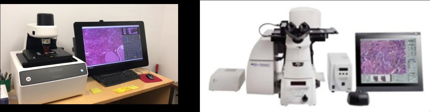
Complex tissues often contain several cell populations packed into the same microscopic region. A protein signal detected across an entire section may therefore reflect multiple cell types at once. Laser capture microdissection services can help researchers isolate selected cells or defined tissue areas so downstream molecular testing begins with a more focused sample.
The challenge is deciding exactly which cells should be collected. Morphology may provide enough guidance in some studies, but biomarker expression can offer a more precise roadmap when visually similar cells have different biological roles.
Use Protein Localization as a Spatial Guide
Researchers can first examine marker distribution across tissue to identify regions with distinct protein expression. This creates a spatial reference that can be compared with morphology, anatomical boundaries, and the experimental question before any material is removed.
Well-designed immunohistochemistry kits support this process by enabling antibody-based detection of selected proteins in tissue sections. When marker localization is reliable, investigators can use that information to distinguish populations that might otherwise be difficult to separate by appearance alone.
Plan Marker Selection Carefully
A useful marker should answer a specific question. Researchers may want to identify epithelial cells, stromal populations, immune infiltrates, vascular structures, or another compartment before selecting an area for downstream analysis.
Marker choice should also consider specificity, expected localization, tissue type, and fixation history. A strong signal is not automatically useful if it appears in several unrelated cell populations or if background staining makes boundaries difficult to interpret.
Connect Staining With Microdissection
Immunoguided microdissection can link protein localization with targeted sample collection. Instead of collecting a broad region, researchers can use marker-positive areas as a guide for isolating a population that is more closely connected to the biological question.
This can be valuable in heterogeneous specimens where neighboring cells may have very different molecular profiles. The approach helps reduce dilution of cell-specific DNA, RNA, or protein signals by material from surrounding tissue.
Protect Molecular Quality During Preparation
Staining conditions used before microdissection should be compatible with the downstream assay. Long procedures, harsh reagents, or unnecessary exposure to aqueous solutions can reduce the quality of nucleic acids or proteins in small captured samples.
When RNA analysis is planned, nuclease-free handling becomes especially important. Researchers should use clean equipment, appropriate solutions, controlled staining times, and storage conditions that help preserve the material from sectioning through collection.
Match Collection Size to the Assay
Greater spatial precision often means less starting material. Capturing a very small population can improve purity, but it may also produce DNA, RNA, or protein amounts close to the lower limit of a downstream method.
Researchers should therefore balance enrichment with yield. PCR, sequencing, transcript analysis, and proteomic workflows have different input requirements, so collection targets should be set before the first section is prepared.
Use Adjacent Sections Strategically
One practical approach is to use adjacent sections for complementary tasks. A reference slide can show morphology or biomarker localization, while a nearby section is reserved for microdissection under conditions optimized for molecular preservation.
Because consecutive sections are not perfectly identical, researchers should document orientation and anatomical landmarks carefully. This makes it easier to match regions and reduces the risk of collecting a neighboring area that does not represent the intended population.
Include Controls at Both Stages
Controls are needed for the staining step as well as the downstream molecular analysis. Positive and negative staining controls help confirm that marker localization is credible before it is used to guide tissue selection.
Molecular controls and replicates then help evaluate extraction and analytical performance. Keeping these control strategies connected strengthens confidence that a detected difference reflects biology rather than staining variation, collection error, or low-input technical noise.
Build Reproducibility Into Region Selection
Region selection can become subjective when several researchers review the same tissue. Clear criteria should therefore define which marker pattern, anatomical boundary, or morphological feature qualifies an area for collection.
Digital slide images, annotated regions, sample maps, and collection records can support consistency. These records are particularly useful in multi-sample studies where the same selection logic needs to be applied across different tissues or experimental groups.
Conclusion
Combining protein localization with targeted tissue isolation can turn a heterogeneous section into a more focused source of molecular information. One method reveals where a marker is expressed, while the other allows researchers to physically enrich the selected region for further analysis.
The strongest workflow connects marker choice, staining conditions, tissue handling, collection size, controls, and downstream testing from the beginning. When these steps are coordinated, researchers can preserve spatial context while generating cell-focused molecular data that are easier to interpret.